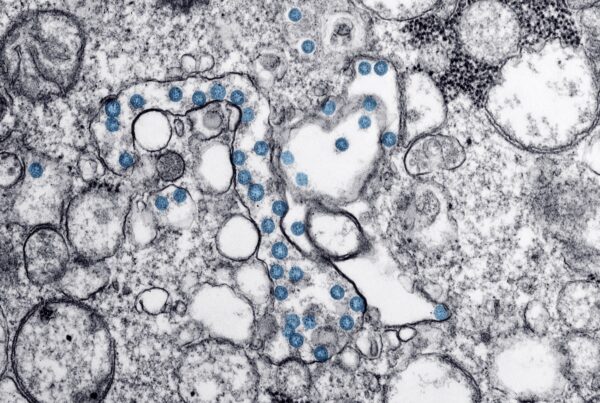
Coronavirus and the GI system: What does the evidence tell us?

The coronavirus continues to have an enormous impact on the way we live. Over the past several months, we have begun to learn more and more about the virus. We’ve learned about how it spreads, its symptoms, how to detect it, and potential approaches for vaccines. Crucially, the virus has several documented impacts and features related to the gastrointestinal system. Let’s take a look at the evidence. 

Note: As the medical and public health understanding of the coronavirus continues to evolve, there’s a lot we still don’t know for certain. It’s important to recognize the difference between evidence-based conclusions, emerging evidence without sufficient peer review, and speculation. We will carefully frame each point based on the amount and kinds of evidence supporting it. 

Coronavirus Symptoms Associated with the Gastrointestinal System

The coronavirus can cause a range of symptoms, from a fever to a loss of smell and a headache. We do know that it can cause gastrointestinal symptoms in some cases. These well-documented GI symptoms include loss of appetite, nausea, vomiting, and diarrhea. However, not everyone with coronavirus will experience these symptoms. 

Some interesting emerging research suggests that there may be clusters of symptoms. One of these clusters involves gastrointestinal symptoms. Researchers at King’s College London studied data from around 1,600 COVID-19 patients who logged their symptoms to the research group’s COVID Symptom Study app in March and April. They found six distinct clusters of symptoms from the data: 

  1. Flu-like with no fever. Headache, loss of smell, muscle pains, cough, sore throat, chest pain, no fever.
  2. Flu-like with fever. Headache, loss of smell, cough, sore throat, hoarseness, fever, loss of appetite.
  3. Gastrointestinal. Headache, loss of smell, loss of appetite, diarrhea, sore throat, chest pain, no cough.
  4. Severe level one, fatigue. Headache, loss of smell, cough, fever, hoarseness, chest pain, fatigue.
  5. Severe level two, confusion. Headache, loss of smell, loss of appetite, cough, fever, hoarseness, sore throat, chest pain, fatigue, confusion, muscle pain.
  6. Severe level three, abdominal and respiratory. Headache, loss of smell, loss of appetite, cough, fever, hoarseness, sore throat, chest pain, fatigue, confusion, muscle pain, shortness of breath, diarrhea, abdominal pain.

As you can see, the gastrointestinal cluster includes GI symptoms like diarrhea and loss of appetite. Other clusters also contain GI symptoms. Each cluster was also associated with differing proportions of patients who required hospitalization and breathing support. The escalating severe clusters had the largest percentage of patients requiring significant medical support. 

It’s important to note that while the researchers have replicated their findings with a second 1,000 person data set from June, the research is not peer-reviewed yet. However, this is certainly something to keep an eye on as more data trends emerge. 

Sewage: A Potential Method for Measuring Infection Spread

Early on in the pandemic, evidence emerged from multiple studies showing that people infected with the coronavirus shed viral particles in their poop. Whether the disease can spread through feces is still undetermined. However, there has been growing interest in sampling sewage to determine the extent of disease spread. 

This has been particularly appealing because of the lack of adequate testing in many countries. This has led to a significant undercounting of actual infection numbers. A cross-sectional CDC study conducted across multiple states used serological testing on a convenience sample with people of all ages to identify how many had developed antibodies to the disease. This gave a more accurate picture of how many people have been infected, since the presence of antibodies indicates that a person had been or was currently infected. The study found that actual infection numbers were likely 10 to 12 times higher than reported through testing. Note that large-scale seroprevalence studies like this one are continuing to be conducted to identify likely infection rates, so the results here are preliminary. 

With such a disparity between reported and actual case counts, wastewater sampling offers a convenient and accessible way to identify disease spread in a particular area. Importantly, results are quicker than those from viral and serological tests. This is helpful, timely data that can inform decisions about disease containment in the event of a flare-up. Additionally, it can help researchers see the viral ancestry of the disease, tracking different strains, viral changes over time, and spreading patterns and paths. 

There are drawbacks, though. This kind of sampling can’t prove that an entire population is completely clear of the virus. Another drawback is that we still don’t know how many copies of viral RNA need to be present in a sample for disease to be detected. This means false negatives are possible, as we don’t know the minimum number of copies that trigger detection. A lower count of copies could go undetected with an improper assumption about a minimum. 

Overall, there’s certainly a lot we still have to learn about the coronavirus and the GI system. However, as scientists conduct more and more research, we have a better opportunity to make data-informed decisions at public health, medical, governmental, interpersonal, organizational, and individual levels. 

Our experienced team at GHP has years of experience helping patients with a variety of diseases and conditions. We can help establish the best plan of care for your situation. Contact any of our office locations to learn about the options we offer and schedule an appointment today.

Endoscopic Retrograde Cholangiopancreatogram (ERCP): A Brief Overview

An Endoscopic Retrograde Cholangiopancreatogram (ERCP) is a therapeutic endoscopic procedure that helps diagnose and treat diseases in the liver, gallbladder, pancreas, and bile system. Here’s what you need to know about the procedure. 

When an ERCP is used

There are several reasons your doctor may perform an ERCP. One common reason is to diagnose and treat gallstones that are trapped in the bile duct. They may also perform an ERCP to identify the source of persistent upper right side abdominal pain, to identify a cause of pancreatitis, or to relieve bile duct obstruction caused by tumors. Often, doctors will inject a dye in the bile ducts during an ERCP to assist with X-ray imaging.

Preparing for an ERCP

There are a few important steps to prepare for an ERCP. You will need to discuss any medications you are taking with your doctor. Blood-thinning medications like Coumadin (warfarin), Eliquis (apixaban), Lovenox (enoxaparin), Plavix (clopidogrel), Pradaxa (dabigatran), and Xarelto (rivaroxaban) are especially important to discuss. These can increase the risk of severe bleeding during the procedure, and you should stop taking them before the ERCP. Also, if you use insulin, you may need to adjust timing or dosage on the day of the procedure. Your doctor will also need to know if you have any allergies to medications. Additionally, you will need to fast starting the midnight prior to your procedure. This means avoiding eating from that time until your procedure. 

During the Procedure

Prior to the ERCP procedure, you will go to a pre-op area where nurses will place an IV and take your medical information. You will also speak with an anesthesiologist about the sedation used during the procedure. 

Once you have moved to the procedure room, you will be connected to machines that monitor your vitals during the ERCP. You will be sedated during the procedure. Your doctor will feed the endoscope through your mouth and perform the procedure. The specific techniques and treatments they use will depend on your situation, and include opening blocked ducts and inserting stents. They may also take X-rays during the procedure. Contrast dye is injected through the endoscope to assist in developing X-ray imaging of your bile ducts. In total, the ERCP should take around 30-40 minutes. 

After the Procedure

After the ERCP, you will go to a post-op area to recover from sedation and the procedure and will be monitored for complications. Once you have recovered, your doctor will discuss the results with you, though any biopsies will take a few days to return. You should not operate machinery, drive, or make important decisions for 24 hours after your procedure due to sedative effects. 

It’s common to have a sore throat immediately after the procedure; throat lozenges can help treat soreness. You should follow a clear liquid diet after the ERCP, eventually transitioning to bland foods. 

The procedure has a few uncommon risks. Around 7% of patients experience pancreatitis, an inflamed pancreas. This requires hospitalization to rest the pancreas and manage inflammation. In very rare cases, this can be severe and lead to surgery, organ failure, or death. 

In very rare cases, perforation occurs during the procedure. Most of the time, this can be managed with hospitalization, bed rest, antibiotics, and bowel rest. Rarely, you may need surgery to repair the perforation. 

There is a low risk of bleeding as well, which occurs in around 0.5% of cases. When managed during the procedure, it can be stopped. Delayed bleeding requires patients to return to their doctor for treatment. 

Our experienced team at GHP has years of experience performing ERCPs. We can help establish the best plan of care for your situation. Contact any of our office locations to learn about the options we offer and schedule an appointment today.


Endoscopic Ultrasound: How to Prepare

An Endoscopic Ultrasound (EUS) is a procedure for assessing and producing images of the digestive system with an endoscope. It is also used as a modality to treat certain gastrointestinal disorders through fine-needle aspiration (FNA). Follow along for an overview of EUS. 

When an Endoscopic Ultrasound is used

An EUS can help doctors determine the source of chest pain, abdominal discomfort, and other symptoms. It can also help them evaluate the extent of disease spread in your digestive tract, and evaluate findings from other diagnostics like MRIs or CT scans. It can help evaluate conditions including Barrett’s Esophagus, Lymphoma, and various cancers. 

There are many different therapeutic procedures that can be performed during an EUS. These include celiac plexus neurolysis (EUS-CPN), pseudocyst drainage, biliary drainage (EUS-BD) and liver biopsy. Each of these treatments has a unique set of steps involved. Below, we will take a look at some general steps and approaches involved with the various diagnostics and treatments performed through an Endoscopic Ultrasound. 

Preparing for an EUS

The way you need to prepare for an EUS depends on the specifics of the procedure and any therapeutic treatments in your case. In general, you often need to fast starting the midnight before the procedure. This helps empty your stomach. You may also need to discuss any medications you are taking with your doctor before the procedure. It is especially important to stop taking any blood-thinning medications leading up to the procedure. These medications can increase the risk of severe bleeding during the Endoscopic Ultrasound. If the EUS is being performed in the rectal area, you may need to undergo a cleansing routine to prepare your bowels. This usually consists of taking a liquid laxative and sticking to a liquid diet for a day before the procedure. 

During the procedure

On the day of your Endoscopic Ultrasound procedure, you will first go to a pre-op area where your medical information will be taken and you will be given an IV. You will also discuss sedation used during the procedure with an anaesthesiologist. 

Depending on the location in your GI tract that doctors will examine or treat, they will advance an endoscope through your mouth or a colonoscope through your anus. You will be on your left side and may be sedated as this happens. Your doctor will advance the scope to the site or sites of interest and then perform any diagnostics or treatments. This can include tissue biopsies, pain-relieving injections, pseudocyst drainage, and bile duct drainage, depending on your situation. Your doctor will be able to see your GI tract through a camera at the end of the scope, and will pass any necessary instruments through the scope to perform the procedure. Most EUS examinations take under an hour, but the exact time will vary depending on what diagnostics or treatments your doctor is administering. 

After an Endoscopic Ultrasound

After your procedure, you will go to a post-op area where you will recover from any sedation and be monitored for side effects of the procedure. Once you are recovered, your doctor will share results with you. Some biopsy results may take longer to return. Given sedation side effects, you should not operate machinery, drive, or make important decisions for 24 hours following the procedure. 

Endoscopic Ultrasounds have a relatively low risk for complications. If you underwent Celiac Plexus Neurolysis to provide pain relief for tumors, you may experience abdominal pain for a few hours and diarrhea for a few days. More rarely, bleeding, infection, and paralysis can occur. If you underwent Pseudocyst Drainage, there is a small risk of bleeding, infection, and pancreatitis. Additionally, in under one percent of cases, perforations occur, requiring surgery to repair. If you underwent Biliary Drainage, there is a 10-20% chance of mild complications associated with bile drainage. These include bleeding, infection, and bile leakage in the abdominal cavity. If you underwent a Liver Biopsy, complications are very rare, and include a small risk of bleeding and infection. In general, if you were sedated during your procedure, there are a few uncommon complications including aspiration, adverse reactions to sedative medication, and complications from lung and heart diseases. 

Our experienced team at GHP has years of experience performing Endoscopic Ultrasounds. We can help establish the best plan of care for your situation. Contact any of our office locations to learn about the options we offer and schedule an appointment today.

Endoscopic Mucosal Resection (EMR): An Overview

Endoscopic Mucosal Resection, or EMR, is a therapeutic technique that helps remove precancerous and early stage cancer lesions during an upper endoscopy or colonoscopy. This technique is a less-invasive alternative to surgery. Here’s what you need to know if you are preparing for an EMR.

When is an Endoscopic Mucosal Resection Used?

EMRs are used to remove abnormal tissues in the digestive tract. The procedure can help treat a variety of conditions including Barrett’s Esophagus, colorectal cancer, and colon polyps. This is a less invasive option than surgery for removing abnormal tissues in the digestive tract. While EMRs are often used to treat disease, your doctor may also collect tissue samples during the procedure. They can examine tissue they collect to determine a diagnosis and the scope of disease spread. 

Preparing for an EMR

There are several important steps you need to follow prior to an Endoscopic Mucosal Resection. Your physician will discuss each of these with you leading up to the procedure. First, you may need to stop taking certain medications. These include blood-thinning medications like aspirin, Plavix (clopidogrel), Xarelto (rivaroxaban), Lovenox (enoxaparin), Pradaxa (dabigatran), Coumadin (warfarin), and Eliquis (apixaban). Blood-thinners increase your risk of excessive bleeding during the procedure. Also, if you use insulin, you may need to adjust your dosage and timing before the procedure. Make sure you discuss all medications you use with your doctor. 

Additionally, you will need to follow a clear liquid diet the day before the procedure, and stay hydrated. You will also need to fast beginning the midnight before your procedure. For EMRs performed through a colonoscopy, you will also need to undergo a cleansing routine. This involves taking a liquid laxative the day before your EMR to prepare your bowels. 

During the procedure

EMRs are performed through either an upper endoscopy or a colonoscopy, depending on the location of the diseased tissue. When you arrive for your procedure, you will go to a pre-op area where nurses will take your medical information and place an IV. You will also speak with an anaesthesiologist about the sedation they will use for the procedure. 

You then will go to the procedure room and be connected to monitors that will measure your vitals during the EMR. You’ll be sedated at this point. If the procedure is done through an upper endoscopy, you will be placed on your left side and given a bite block so the endoscope can pass through your mouth safely. If it is done through a colonoscopy, you will also be placed on your left side so the colonoscope can pass through your anus and advanced into the colon. 

Your doctor will be identifying and removing lesions during the EMR. There are several ways to remove lesions. Your doctor may inject a liquid into the submucosal layer under the lesion, which acts as a pillow that lifts the lesion for easy removal. They may also use a suction or a rubber band to help lift the lesion. After the lesion is lifted, it will be captured with a snare and the removal site will be cauterized. The procedure takes around 20 to 60 minutes.  

After the Endoscopic Mucosal Resection

Following your EMR, you will move to a post-op area to recover from sedation and monitor for any complications. Once you have recovered, your doctor will talk to you about the findings and give you post-op recovery instructions. You should not drive or make important decisions for 24 hours following the EMR due to sedative effects. You should follow a clear liquid diet immediately following the procedure, and can later transition to bland foods and a more regular diet. 

Complications from an EMR are uncommon. This includes bleeding, which occurs in 5-10% of cases. Your doctor can usually stop bleeding during the procedure if they recognize it. However, bleeding can become severe if it is delayed and may require follow-up care. In other rare cases (1-2% of the time), perforation of the intestine can occur. This is often managed through antibiotics, bowel rest, and hospitalization. It may require surgery as well. Additionally, some patients have reactions to sedative medication in uncommon cases. Always contact your doctor if you have any severe symptoms like abdominal pain, a fever, or excessive rectal bleeding after the procedure, as they may indicate a severe complication. 

Our experienced team at GHP has years of experience performing EMRs. We can help establish the best plan of care for your situation. Contact any of our office locations to learn about the options we offer and schedule an appointment today.